
25-03-2026 10:35
Hulda Caroline HolteHello,I collected this species growing on a dead b
26-03-2026 15:31
 Åke Widgren
Hello,I found this one in October last year, on r


25-03-2026 22:23
 Marc Detollenaere
Dear Forum,On a debarked stem of Tilia, we found s
24-03-2026 15:44
Åge OterhalsI hope someone can confirm the name of this collec
25-03-2026 20:53
François BartholomeeusenDear forum members,On 23 March 2026, I found sever
23-03-2026 20:16
 Miguel Ángel Ribes
Good eveningI'm unable to identify this Coprotus o
25-03-2026 15:06
 Bernard CLESSE
Bonjour à toutes et tous,Pourriez-vous me confirm
25-03-2026 13:54
 Enrique Rubio
Does anyone know where I could download Paoletti's
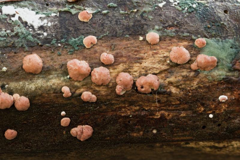
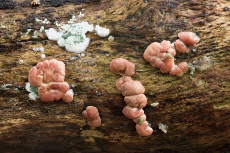
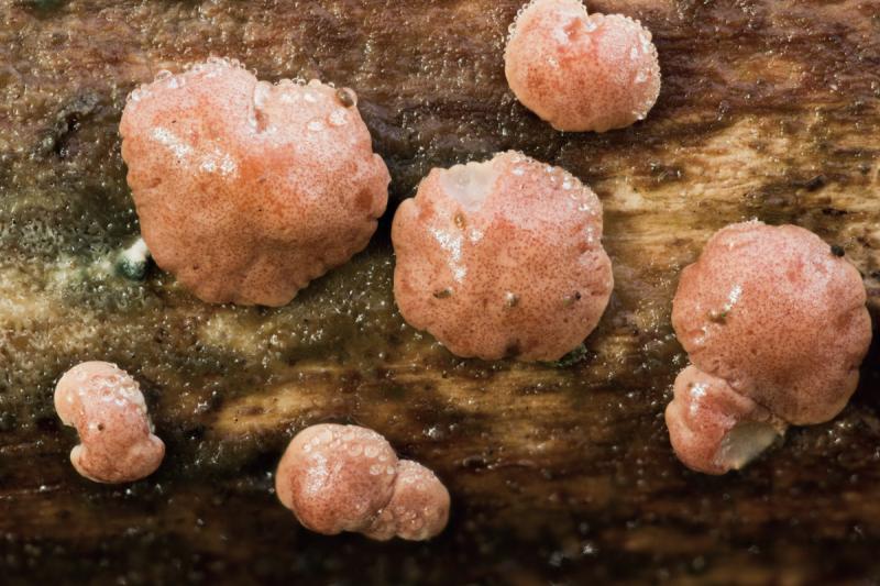
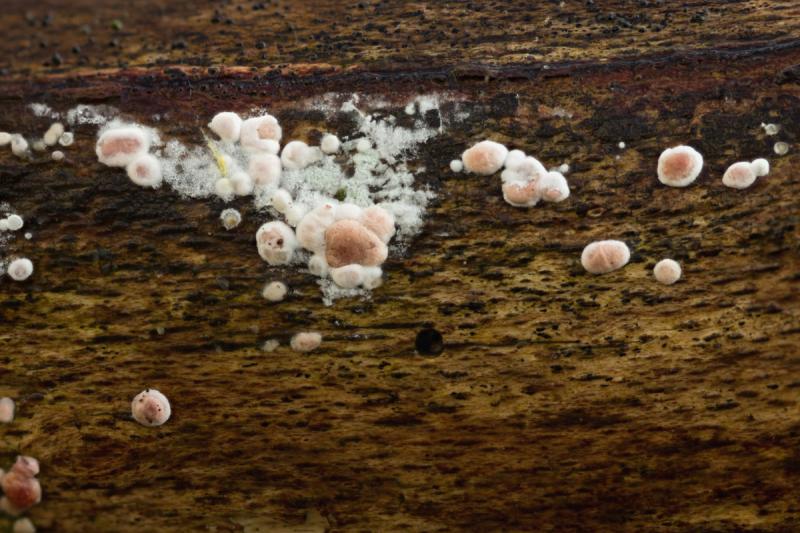
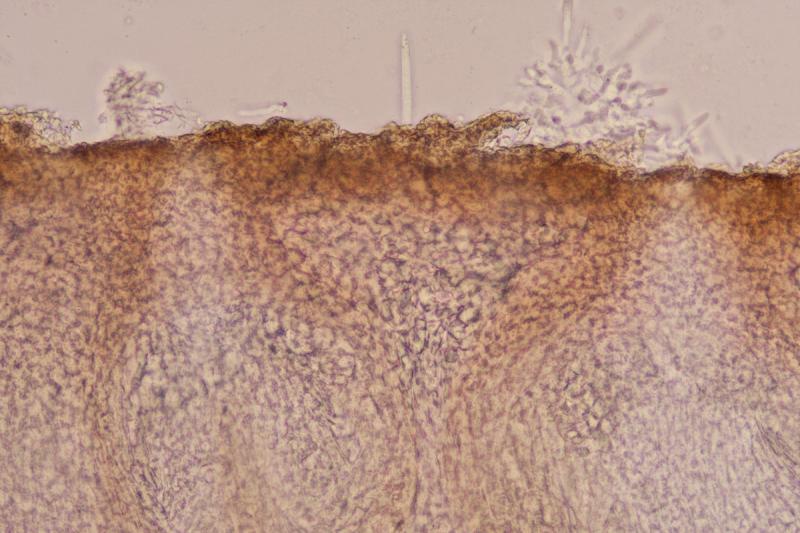
Here is what I believe is a Hypocrea found on Fagus branch on the forest floor. The stromata are not mature but the green anamorph was present which may help to identify it. The stromata were to 5mm and there were many of them. Attached to the substrate right up to the edge. They were a pinkish colour with visible darker ostiolar openings creating a pattern of dots on the surface. They dried to a pale brown.
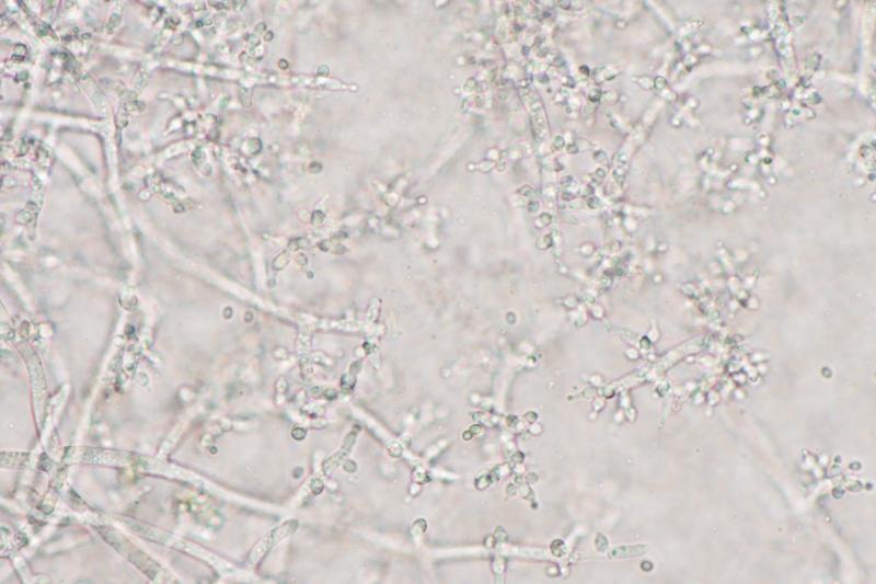
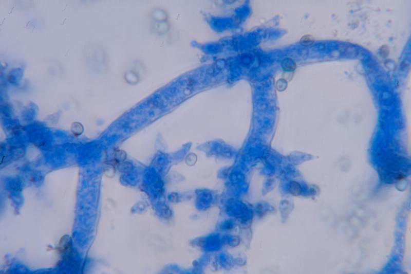
The anamorph was green and I don't have the language to describe the structure of the conidiophores and phialides, hopefully the pictures will help
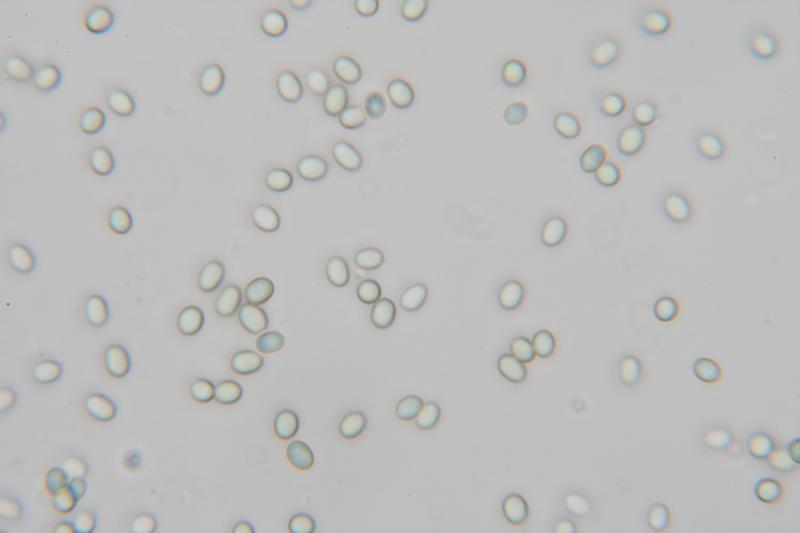
The conidia were smooth and ellipsoid 3.3 X 2.4 QE 1.4
My attempts to key it give Hypocrea minutispora and that does look OK with the structure of the conidiophores and phialides as described for that species in this paper although the conidia may be a bit on the large size.
If anyone can help I would appreciate it.
Many thanks
David

I think your fungus is H. minutispora but it would be necessary to compare it to H. pachybasidioides.
Christian
Thank you Christian,
I looked again at the paper I mentioned in my first post and it appears that H. pachybasidioides is in the "polysporum" clade and its anamorph, (Trichoderma Polysporum) has white/hyaline conidia where my specimen had green conidia. It looks like it also has infertile, corkscrew-like extensions of the conidiophores unlike my specimen. The green, smooth, ellipsoid conidia, along with the structure of the conidiophores pictured in the paper above, do seem to leave only T. minutisporum / H. minutispora.
Regards
David