
12-06-2026 14:50
 François Freléchoux
Bonjour, Voici la brève description d'une Mollis
10-06-2026 21:16
François Freléchoux
Bonsoir,Le dernier du jour, en attendant votre avi
11-06-2026 19:01
 William Slosse
Hello all,In an attempt to make a culture of a sus
11-06-2026 19:03
 Nicolas VAN VOOREN
Chers membres d'Ascofrance,Le site sera placé en

09-06-2026 18:32
Camille MertensSur morceau de roseau immergé 0,5 - 0,7 mm de dia
10-06-2026 12:54
Steve ClementsBonjour encore, Pouvez-vous m'aider, s'il vous pl
10-06-2026 21:07
François Freléchoux
Toutes les tiges de gentianes jaunes de l'an pass�
10-06-2026 13:41
François Freléchoux
Bonjour à nouveau, Voici une trouvaille d'hier.
previous post of Exarmidium diaphanum gave me a hint about the position of one my unidentified specimen,
it was collected on dead branch of Chamaedaphne calyculata (N61,063892° E69,455695°). There is, probably, candidate, E. ericae, which described from twigs of Erica carnea (the same family) - i am comparing it now.
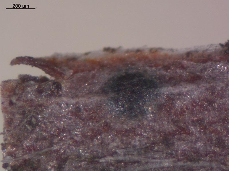
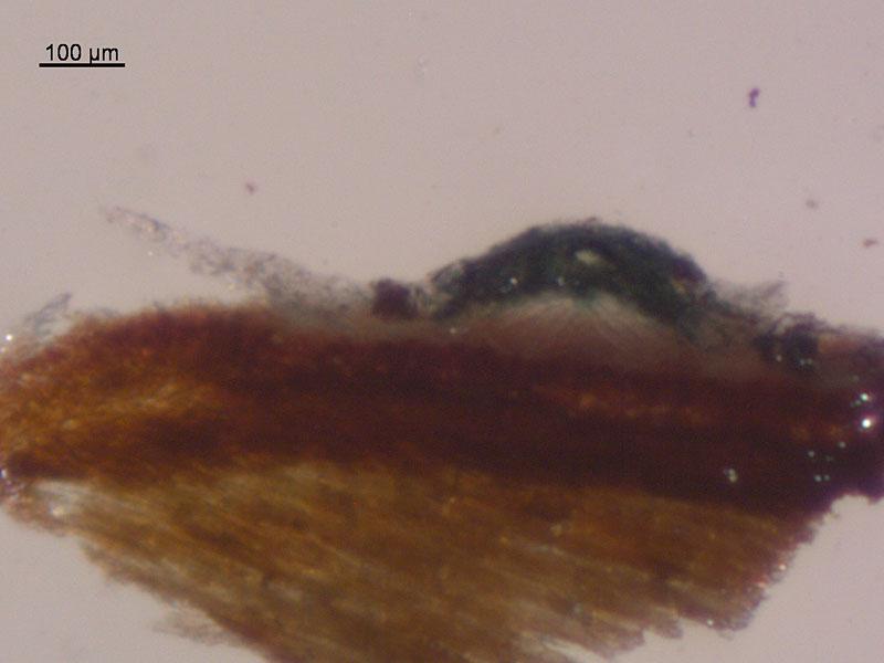
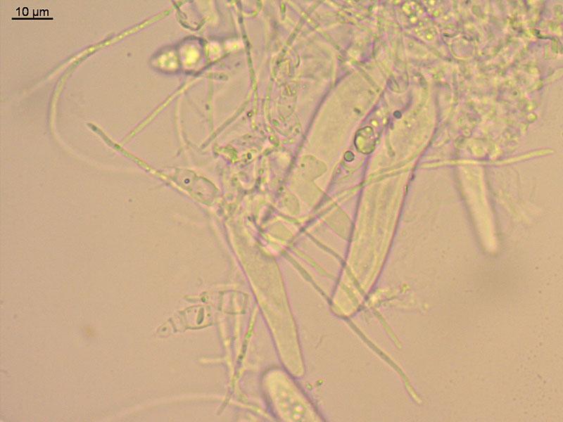
Hymenium developing under the clypeus, flat, with tiny pore (30 mk), to 400 mk broad, 80 mk thick, clypeus bluish-brown.
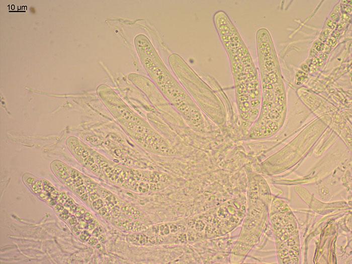
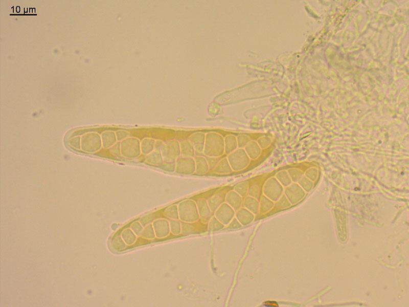
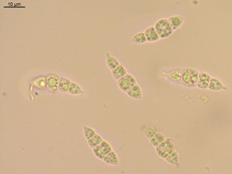
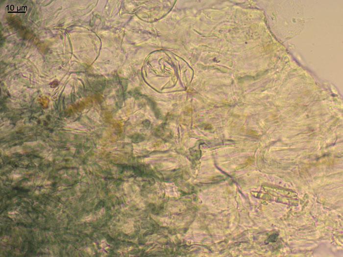
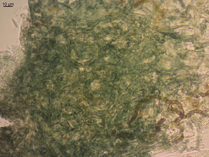
Clypeus from host epidermis cells and mass of greenish-blue chains of cells (some symbiont), no fungal tissue presented in clypeus (? may be it is not right to say clypeus then); asci fissitunicate, not amyloid, enlarged at base and with obtuse tip, thick-walled apex, 76 (70-85) x 16 (14-17); paraphyses cylindrical, 1,3-1,5 mk thick, with some guttules, branched; spores fusoid, 3-4-celled, cells of released spores enlarged to round (and then overall shape with constrictions), with two short cylindrical outgrowths from both ends, content with several big and many small guttules, 23,7 (20,8-26,6) x 7,9 (6,4-9,2) (18 spores).
sorry, but I can't help you with this one.
But thank you for reminding of unfinished determinations:
http://www.ascofrance.fr/search_forum/11375?
I should keep this in mind next spring-summer.
Best wishes for your determination: Marja