
18-05-2026 12:43
Sylvie Le GoffBonjour à tousPuis je avoir votre aide sur ce que
19-05-2026 12:55
 Hardware Tony
After checking Gminder and Otto's library I cannot
19-05-2026 10:27
Patrice TANCHAUDBonjour, récolte récente sur terre retournée i

18-05-2026 10:13
 Lieve Deceuninck
Dear forum members,I identified this as the teleom

17-05-2026 19:05
Thomas FlammerI have found this tiny 200 ym cup shaped apothecia
17-05-2026 16:41
Margot en Geert VullingsWe found this Lachnum on an old Rubus stem.Fruitbo
05-04-2026 22:46
 Lothar Krieglsteiner
on wood of Ceratonia, Algarve, 3.4.2026.The color
15-05-2026 13:33
Sylvie Le GoffBonjour à tousJe serais très reconnaissante enve
Eutypa? Cryptosphaeria?
Björn Wergen,
17-01-2011 13:25
 Hello,
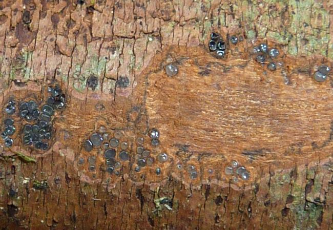
I have collected a Diatrypaceae on ?Prunus spinosa, perhaps the substrate is wrong and you can help me with determining this, I am at least not really sure with determining Crataegus and Prunus in winter :D
well, here are some features of this species:
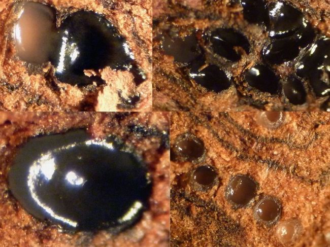
Fruitbody consisting of single or a bit clustered perithecia completely embedded in the cortex, 0,2-0,3 mm, first withish-greyish, then black, with a rudimentary ostiolus (not easy to see), after cutting with black lines around the fruitbody (rudimentary stroma?).
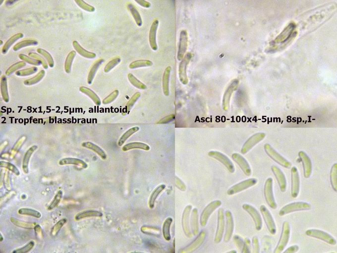
Spores 7-8x1,5-2,5 µm, allantoid, pale brownish, with 2 little vakuoles in the endings, smooth. Asci 80-100x4-5 µm, long stalked with clustered spores in the middle, with 8 spores (biseriate), IKI-, Paraphyses hyaline, with a few vakuoles, inconsiderable.
It seems as if the asci collapse at maturity (?).
Can someone help me with this? Or at least with literature to determine diatrypaceae? I have always problems with this ;)
regards,
Kazuya
Björn Wergen,
17-01-2011 13:27
Re:Eutypa? Cryptosphaeria?
photo2 ca. x40
Björn Wergen,
17-01-2011 13:28
Re:Eutypa? Cryptosphaeria?
micro
Jacques Fournier,
18-01-2011 18:11

Re:Eutypa? Cryptosphaeria?
Hi Kazuya,
I think you are indeed dealing with a Diatrypaceae on Prunus spinosa. The rectangular pattern of cracking of the bark seems typical.
On this host Eutypa lata is very frequent but not much typical because it forms small stromata as on your photo, leaving the periderm intact. Two key features of this species are the nearly smooth, non sulcate ostioles, and IKI+ apical ring. This structure is very small and the bluing rather faint, you have to observe it on a very thin slide. You can use either IKI or Melzer's reagent after pretreatment with 3% KOH to stain the apical ring of Diatrypaceae, but I prefer the second method which involves a retraction of the protoplasm in the ascus and makes the minute ring more conspicuous.
Identifying Diatrypaceae is a hard job, not much rewarding until you have some practice. The only comprehensive taxonomic treatment is that of Rappaz 1987. I think you can find a pdf version on Ascomyceteorg.
Good luck!
Jacques
Björn Wergen,
19-01-2011 01:15
Re:Eutypa? Cryptosphaeria?
Hm many thanks jacques, I've already visited the site, there is also the information about Rappaz 1987 and a link but I'm not allowed to see the page without a password...ascomycete.org is new, right? I didn't hear about it before :D
regards,
Kazuya
regards,
Kazuya
Björn Wergen,
19-01-2011 20:50
Re:Eutypa? Cryptosphaeria?
Hey! What about Cryptovalsa protracta?? But with more than 8 spores...:(
Jacques Fournier,
19-01-2011 22:19
Re:Eutypa? Cryptosphaeria?
Cryptovalsa protracta has 32-spored asci, larger spores and the stroma, although higly variable, usually is Eutypella-like, pustulate to pulvinate. It cannot be confused with E. lata!
No idea about your Dothideomycete, you should look into the structure of the "stroma". Try to make a thin section and take a photo under the microscope.
Good evening!
Jacques
No idea about your Dothideomycete, you should look into the structure of the "stroma". Try to make a thin section and take a photo under the microscope.
Good evening!
Jacques