
12-12-2024 09:30
 Josep Torres
Hello.I found some small black perithecia on an oa
03-12-2024 12:47
 Vasileios Kaounas
Hymenoscyphus conscriptus ? In Hedera helix, in f
14-11-2020 20:06
 William Slosse
Good evening all,today 14/11/20 we found the follo
11-12-2024 19:47
 Zoltan Lukacs
Dear Friends, Anybody has a pdf of these works ?
12-12-2024 01:07
 Viktorie Halasu
Hello, would anyone have this paper please? Matz
05-12-2024 16:38
Karl Soler KinnerbäckA Melastiza from Padjelanta, alpine Sweden, on cal

09-12-2024 11:26
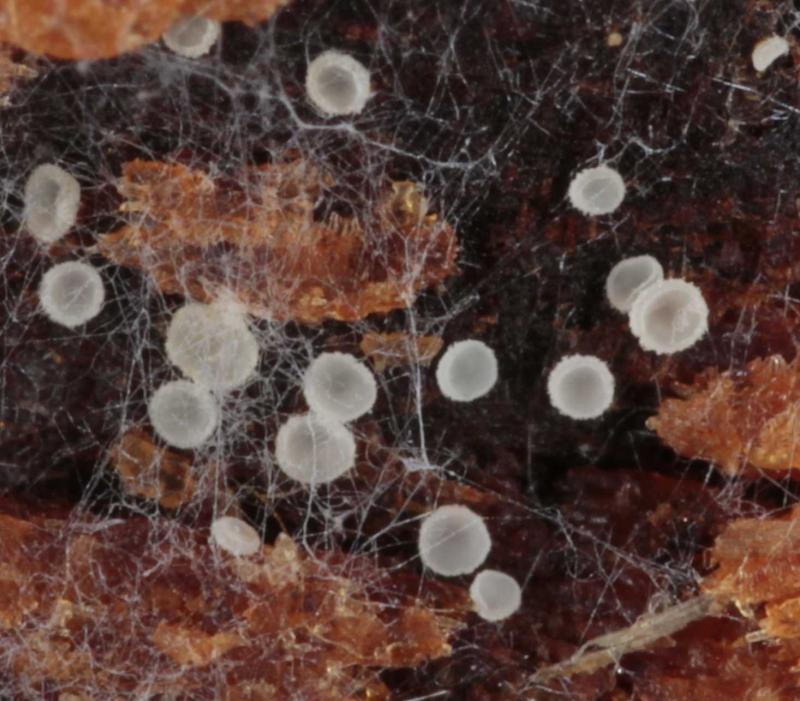
Louis DENYHello forumSur tige morte de Rubus sp. Récolté �
 Rebonjour,
Comme annoncé dans mon post précédent, sur le même tronc, les deux espèces en mélange.
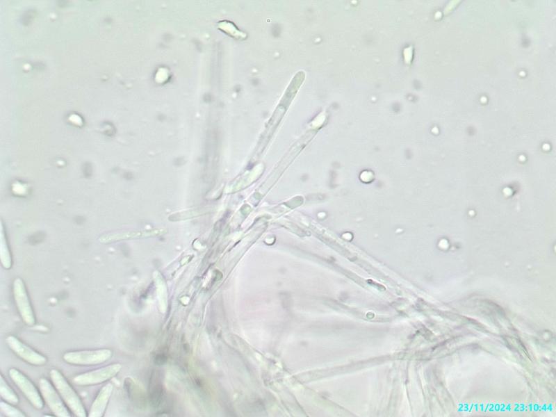
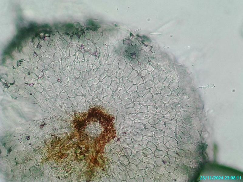
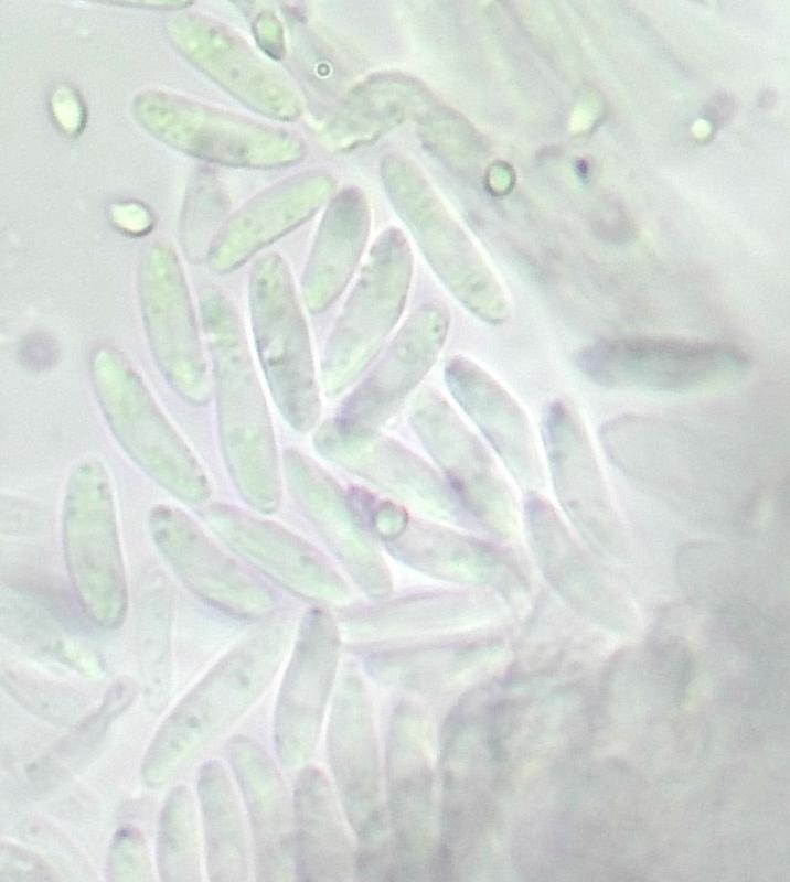
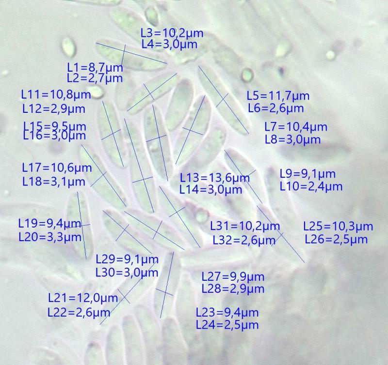
Récolté "dans" un tronc de Picea abies imbu et assez dégradé le 31/10/24 dans les Vosges, alt 850m.
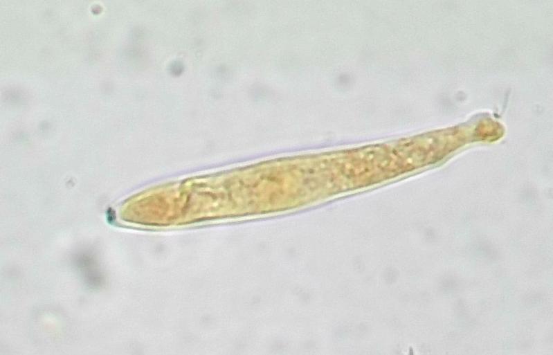
Apothècies cupulées blanches, asques octosporées, bisériés, crochets+ IKI+, paraphyses +/- capitées, poils cylindriques lisses, excipulum ectal à texture prismatica-globulosa, spores 9,5-11,5 X 2,5-3µ, ellipsoides allongées, quelques rares et petites guttules.
Je n'arrive à nouveau pas à m'orienter vers un genre..., merci pour votre aide.
éric.
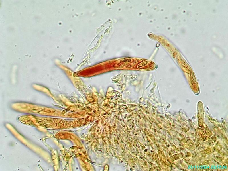
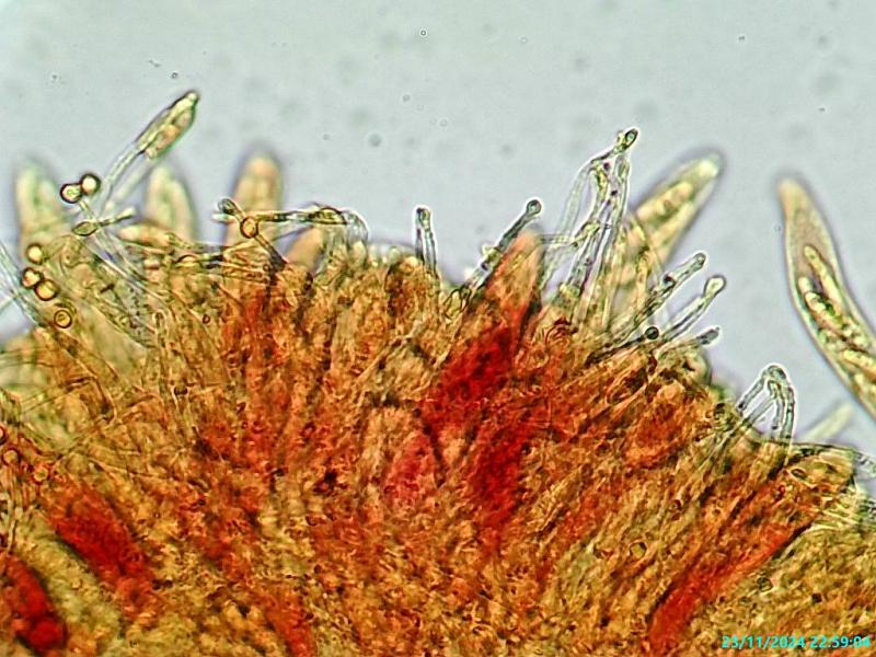

En faisant une préparation de la marge, tu trouverais probablement des poils correspondant au genre Hyaloscypha (mais de grâce, pas dans le rouge congo -:) )
AmitiésMichel