15-07-2014 12:51
Gernot FriebesHi,identifying Lambertella species is difficult en
21-06-2024 18:18
Sylvie Le GoffBonjourpourais je avoir confirmation pour Lachnum
20-06-2024 16:07
 Nicolas Suberbielle
Bonjour, J'ai trouvé cette espèce sur les feuil
19-06-2024 18:32
François BartholomeeusenOn female Alnus catkins I found very small fruit b

16-03-2018 12:26
Garrido-Benavent IsaacDear all,I found this beautful pseudotheciate asco
19-06-2024 17:52
 Bernard Declercq
Dear all, I am looking for the description and ac
18-06-2024 14:07
 Thierry Blondelle
Bonjour,Récolte sur une brindille dans une tourbi
16-06-2024 16:47
 Hardware Tony
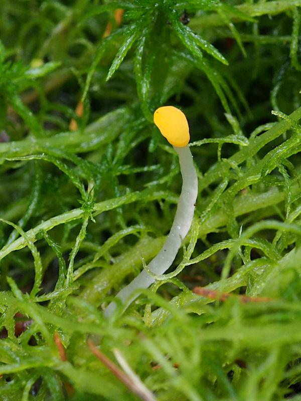
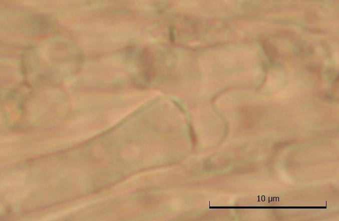
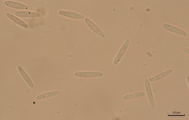
These apothecia directly growing on a resupinate,
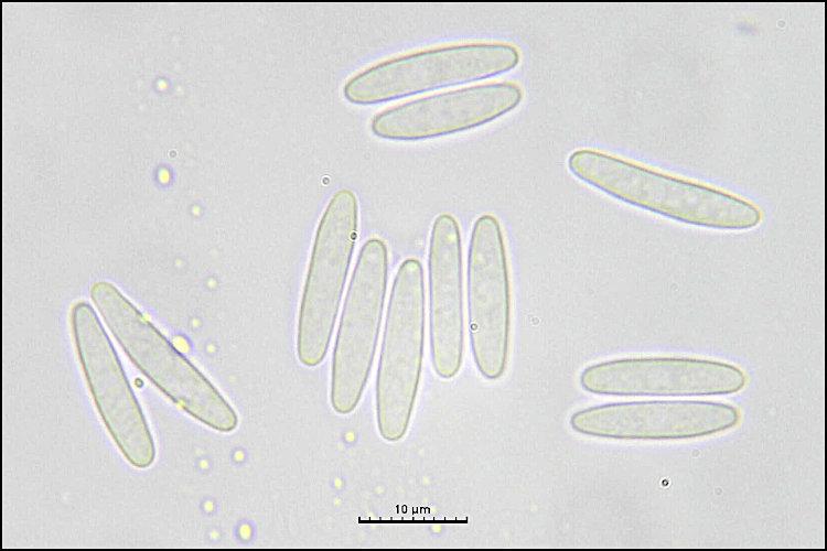
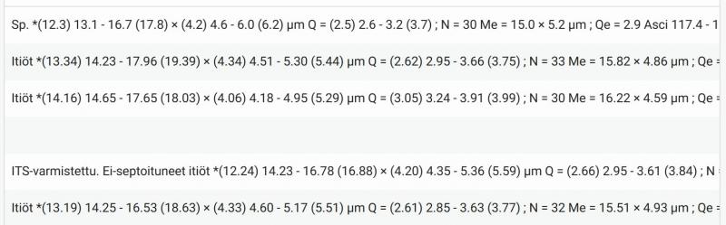
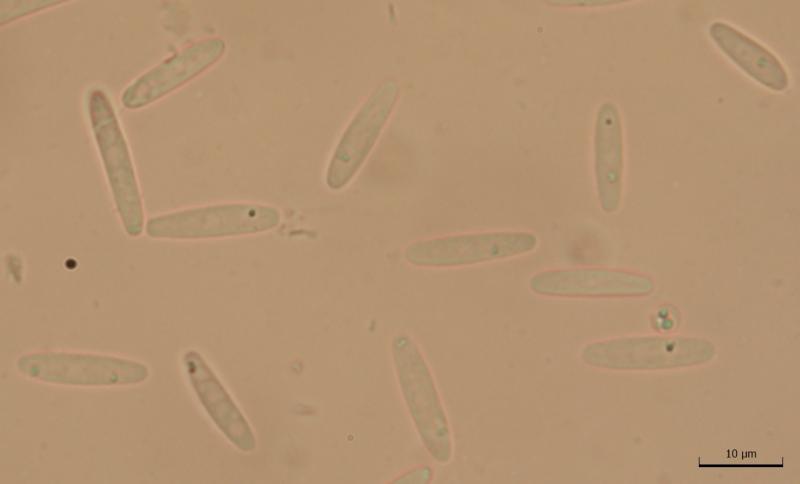
Spore measurements:
(13.5) 14.6 - 17.6 (18.1) × (2.8) 3.2 - 4 (4.2) µm
Q = (3.4) 3.9 - 5.3 (5.8) ; N = 20
Me = 15.9 × 3.6 µm ; Qe = 4.5
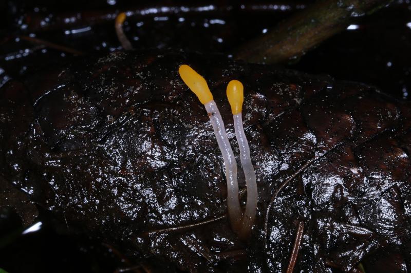
The old paper The genus Mitrula in North America (1977) doesn't seem to clearly key out this specimen. Either this is M. paludosa or M. borealis. The spore measurements would fit M. borealis slightly better, but the spores didn't appear to have a sheath (fresh spore deposit), which might favour M. paludosa.
Any opinions?

Mitrula borealis seems to be very common in SW Finland, perhaps more so than M. paludosa. Some of my observations are recorded here, but I have many more:
https://laji.fi/observation/list?target=mitrula%20borealis
A few weeks ago I collected a Mitrula with spores even broader than borealis according to the Redhead paper. I don't know is it a borealis or still a new one.
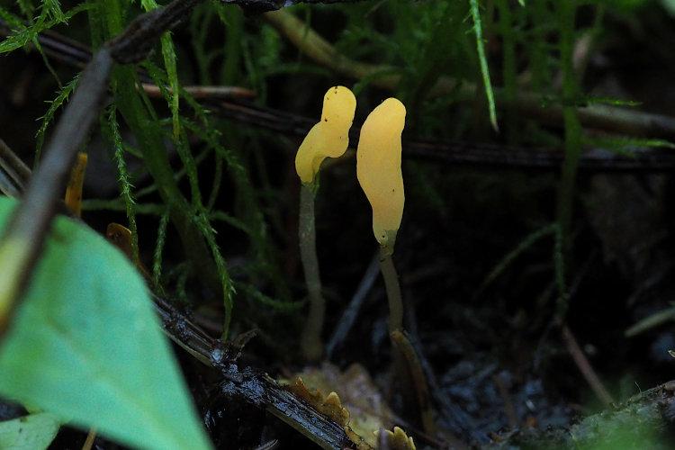
About macroscopic differences I am not sure but the working hypothesis is that borealis is more yellow and perhaps bigger. Paludosa might have more brownish tints in the hymenium and pehaps not so elongated hymenial head as borealis.
Likewise, about ecology I have only a working hypothesis: borealis might be more common in spring fed small streams or pools while paludosa is found in any pool or ditch. All acid, virtually no calacreous sites where I collect.
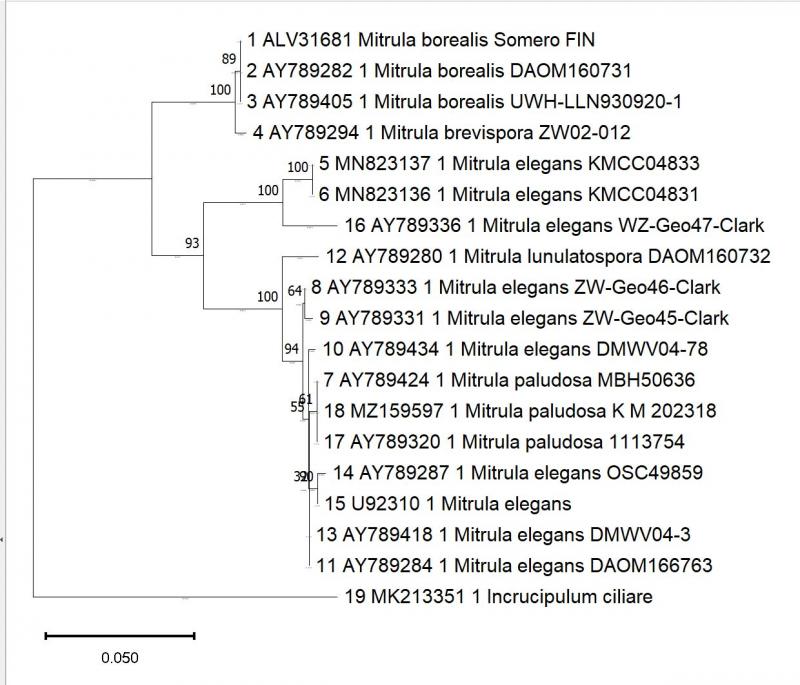
The sequenced one is this:

interesting topic! I must admit I have automatically identified all my Czech collections as M. paludosa. In Czech literature, I haven´t found a mention about M. borealis, but I can see that Redhead mentions occurrence in the neighbouring Germany.
I wonder how often is M. borealis collected in this region...
Yesterday I found Mitrula in the Jeseníky Mts., so I made the first microscopy of it in my life, and it fits better M. paludosa, especially if Redhead worked with dead material. In any case, the are thinner than those in the collections from Finland presented here. Living ascospores measure (13)14.1-18.2(19.2) × (2.8)3-3.6 (3.8) µm, Q = (3.8)4.2-5.6(6.2), n = 40. Me = 15.8 × 3.3 µm; Qe = 4.8. Gelatinous sheath not observed.
Best regards, Zuzana
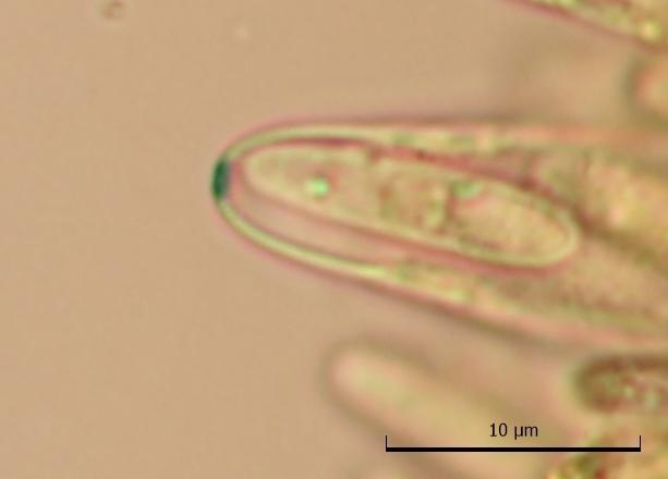
Spores:
(17.3) 18.1 - 21.3 (22.9) × (2.8) 3 - 3.5 (3.8) µm
Q = (4.6) 5.4 - 6.7 (6.8) ; N = 20
Me = 19.6 × 3.3 µm ; Qe = 6