
20-06-2014 08:10
 maurice pelissier
Bonjour trouvé en janvier a Mamoudzou Mayotte à
03-07-2014 17:05
 Nicolas VAN VOOREN
Bonjour.Est-ce que quelqu'un a déjà récolté Wi

03-07-2014 15:56
Salvador TelloHola a todos.Tengo estos hongos creciendo en ramit
02-07-2014 20:22
Gernot FriebesHi,I have a collection of a Tarzetta species here
02-07-2014 15:09
 Uwe Lindemann
Hello Forum, I'm searching for the description of
29-06-2014 13:09
 Rubén Martínez-Gil
Hola a todos. Subo unas fotos de unas Tarzatta qu

Tarzetta 1617
Leandro Sánchez,
03-07-2014 23:33
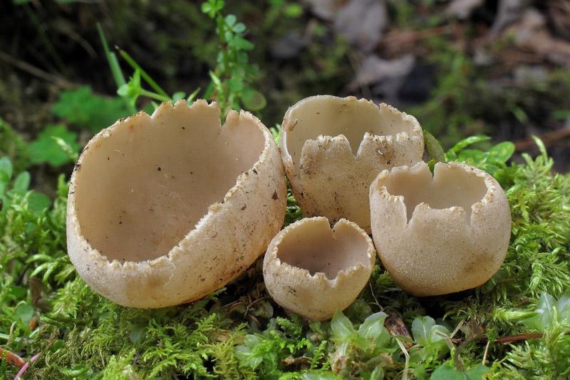
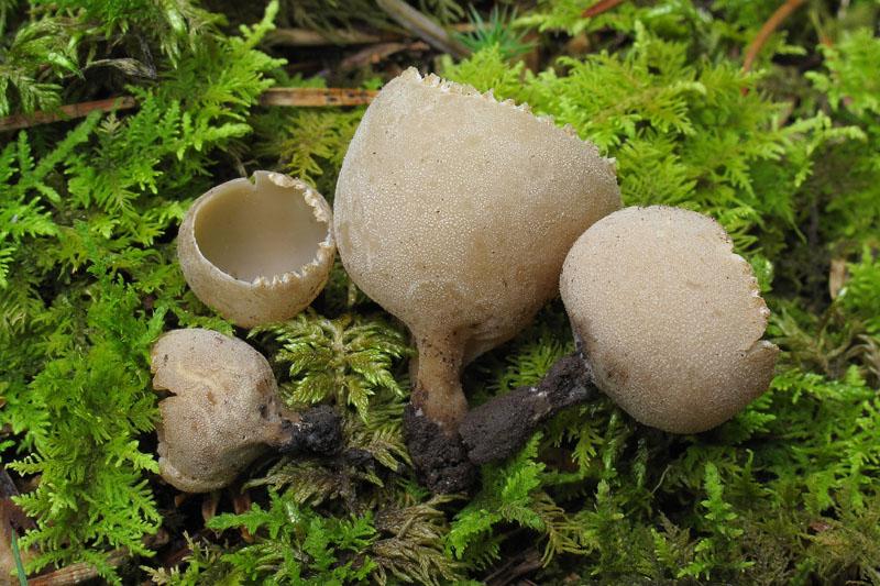
 Jusqu'à 30 mm de diamètre, sous feuillus de rivière et Abies
Asques 240-260/ 11-13
Cordialement
Nicolas VAN VOOREN,
04-07-2014 07:16
Re : Tarzetta 1617
Je pense qu'il s'agit de Tarzetta catinus.
Leandro Sánchez,
04-07-2014 08:16
Re : Tarzetta 1617
Merci beaucoup Nicolas
René Dougoud,
04-07-2014 08:37
Re : Tarzetta 1617
Cher Collègue,
Intéressante récolte !
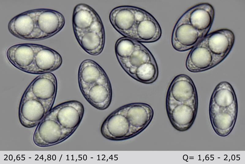
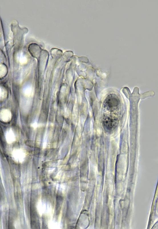
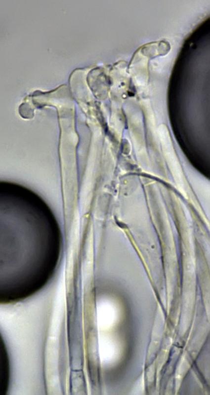
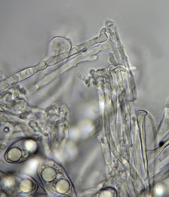
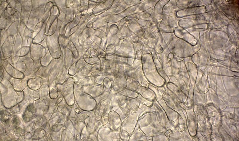
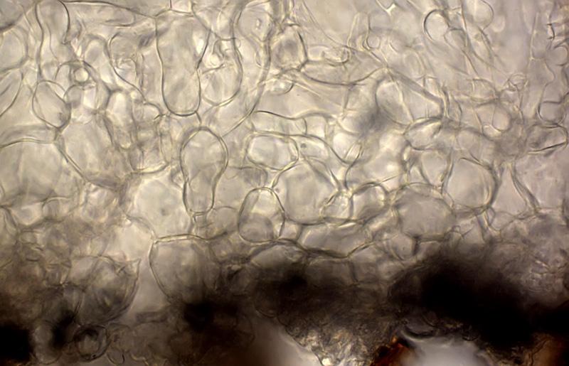
En observant les ascospores de votre récolte (je pars du principe qu'elles ont été photographiées dans l'eau) et en les comparant avec celles de T. catinus (Holm. ex Pers..) Korf & J. K. Rogers, de mes dessins, à la mémoire que j'ai de leur forme et à la photo d'ascospores que présente Ruben-Martinez Gil dans son message du 29.06.2014 sur ce site, je pense qu'il pourrait s'agir de T. spurcata (Pers.) Harmaja. En effet, je relève que les ascospores de votre récolte sont subfusoïdes, soit avec des extrémités amincies, alors que les ascospores de T. catinus possèdent des extrémités plus arrondies. Par ailleurs, et bien que je me méfie de la validité de ce caractère, je relève que les paraphyses de votre récolte sont fortement déformées à leur sommet, ce que décrit Harmaja à la fois pour T. pusilla et pour T. spurcata.
Je souhaite encore relever que T. catinus présente souvent, je dirais même typiquement, soit une perforation ou une déformation au fond de la cupule, ce que montre un des exemplaires de la récolte illustrée par Ruben-Martinez Gil dans son message précité.
De ce qui précède, je pense que votre récolte correspond à T. spurcata Harmaja.
Cordialement
René
Intéressante récolte !
En observant les ascospores de votre récolte (je pars du principe qu'elles ont été photographiées dans l'eau) et en les comparant avec celles de T. catinus (Holm. ex Pers..) Korf & J. K. Rogers, de mes dessins, à la mémoire que j'ai de leur forme et à la photo d'ascospores que présente Ruben-Martinez Gil dans son message du 29.06.2014 sur ce site, je pense qu'il pourrait s'agir de T. spurcata (Pers.) Harmaja. En effet, je relève que les ascospores de votre récolte sont subfusoïdes, soit avec des extrémités amincies, alors que les ascospores de T. catinus possèdent des extrémités plus arrondies. Par ailleurs, et bien que je me méfie de la validité de ce caractère, je relève que les paraphyses de votre récolte sont fortement déformées à leur sommet, ce que décrit Harmaja à la fois pour T. pusilla et pour T. spurcata.
Je souhaite encore relever que T. catinus présente souvent, je dirais même typiquement, soit une perforation ou une déformation au fond de la cupule, ce que montre un des exemplaires de la récolte illustrée par Ruben-Martinez Gil dans son message précité.
De ce qui précède, je pense que votre récolte correspond à T. spurcata Harmaja.
Cordialement
René
Nicolas VAN VOOREN,
04-07-2014 08:48
Re : Tarzetta 1617
Effectivement, les spores paraissent plus "fusoïdes" que l'autre récolte de T. catinus présentée dans un autre sujet. Je trouve également les spécimens un peu pâles pour T. spurcata (= Pustularia ochracea).
Le point que tu soulèves René concernant les paraphyses est intéressant, car j'ai du mal à évaluer la valeur de ce caractère. J'ai quelques collections où on trouve des paraphyses bien droites et d'autres très tortueuses...
Pas toujours facile à déterminer ces Tarzetta :o)
Le point que tu soulèves René concernant les paraphyses est intéressant, car j'ai du mal à évaluer la valeur de ce caractère. J'ai quelques collections où on trouve des paraphyses bien droites et d'autres très tortueuses...
Pas toujours facile à déterminer ces Tarzetta :o)
René Dougoud,
04-07-2014 10:19
Re : Tarzetta 1617
Oui, facile à dire qu'il s'agit d'une espèce du genre Tarzetta, mais bien plus difficile est d'en préciser l'espèce !
S'agissant des paraphyses et de leurs particularités sommitales, il me semble (me basant sur les photos des exemplaires) que la formation de cellules (par bourgeonnement) et/ou leur déformation ne soit pas la conséquence du vieillissement du champignon. Un phénomène que l'on peut en effet remarquer sur certaines discales (operculées et inoperculées) âgées, surmaturées.
Il serait intéressant de pouvoir observer des exemplaires immatures de T. pusilla ou de T. spurcata, afin de pouvoir préciser ou définir si les paraphyses présentes déjà ces particularités sommitales ou à partir de quand ! ?
Il serait également intéressant de savoir si tous les exemplaires de la récolte présentent ce caractère.
Aussi, je demande à notre collègue Sanchez de bien vouloir observer attentivement les exemplaires de sa récolte et de nous informer de ces observations. D'avance merci !
Cordialement
René
Leandro Sánchez,
04-07-2014 10:56
Re : Tarzetta 1617
J'ai observé trois des quatre exemplaires et les paraphyses du exemplaire grand étaient beaucoup plus tortueuses que de jeunes exemplaires
Cordialement
Cordialement