
28-05-2026 16:15
James MitchellHello,Does anyone have the original publication of
28-05-2026 11:06
Thomas Læssøehttps://svampe.databasen.org/observations/10596750
23-05-2026 11:44
 Charles Grapinet
Hello, I am having trouble identifying this copro
25-05-2026 16:44
François BartholomeeusenHi forum members,During an excursion organised by
26-05-2026 21:25
Dirk GerstnerHello everyone, I'm completely stumped by this li
26-05-2026 22:44
Ethan CrensonHi all, I think I have Incrucipulum capitatum her
22-05-2026 14:44
 Lothar Krieglsteiner
in unripe condition citrine yellow, then soon fadi
25-05-2026 16:35
 Bernard CLESSE
Bonjour à toutes et tous,J'ai trouvé récemment,
22-05-2026 13:29
Gernot FriebesHi,I am curious to hear your opinion on this mater
23-05-2026 18:57
Sylvie Le GoffBonjour à tousRécolté sur une branchette de Sal
 • Hymenoscyphus: Habitat, macro, spores, paraphyses.
• H. menthae group: Paraphyses with highly refractive VBs, reddening when traumatised, ectal excipulum with textura prismatica.
• H. menthae: Habitat, simple septa, spores (incl. OCI 5), phenology, although not with the common carotenoids (resulting in whitish colour).
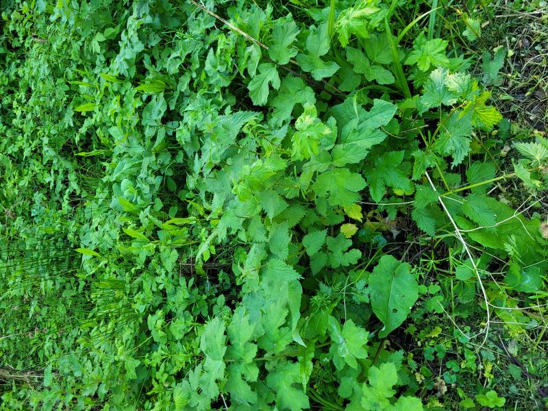
Habitat: On dead, standing stem of Arctium lappa, from last year judging by low level of decay, damp, around the base, close to the floor, completely sheltered by new growth, damp and shady area, two streams joining nearby – often flooding, edge of mixed deciduous woodland, Low Weald, England, early-May.
Associates: Pyrenopeziza cf. atrata (based on macro) on the same part of the stem, brown phragmoconidia found on apothecia, transparent Collembola, small transparent soil mites; in vitro an extensive mycelium from an anamorphic fungus developed - somewhat covering the apothecia of interest, Myxomycete fruiting – cf. Comatricha sp.
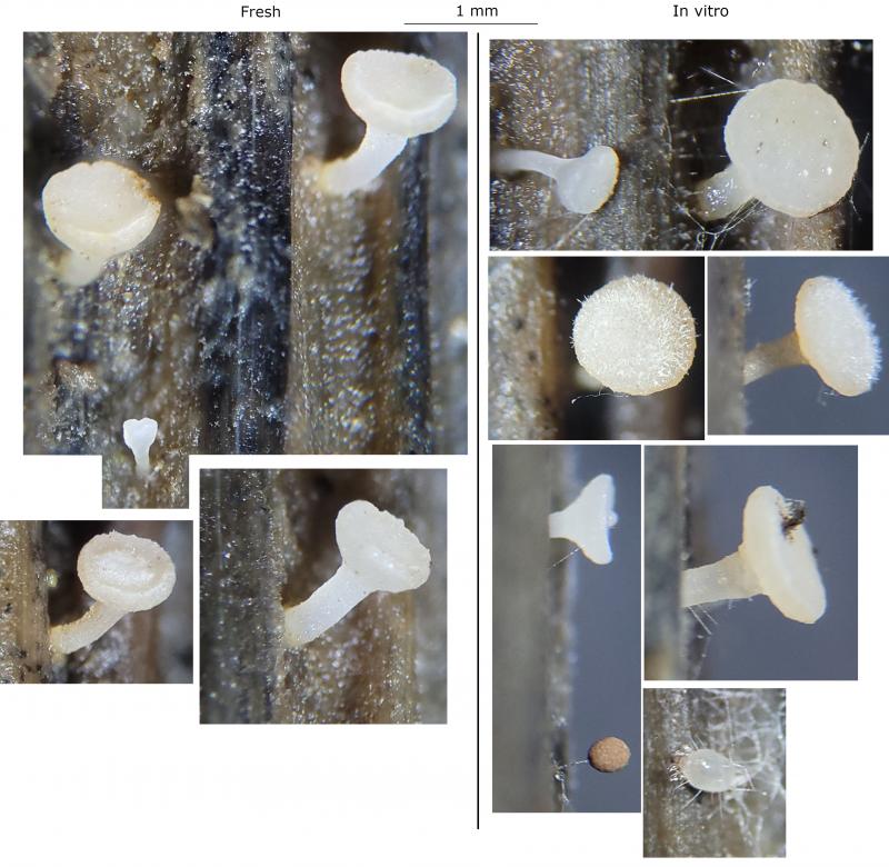
Apothecia (fresh): ~10 on the stem, scattered to gregarious, in several broad groups, mostly appearing superficial but some may have shallowly erupted, 0.6-1 x (0.5) 0.7-1.1 mm, Q ~0.9, whitish-cream, apparently browning with age, stipe developing early, initially cupulate-cyathiform and eventually discoid – giving tack-shape; receptacle more cream coloured, appears uneven but glabrous, somewhat lustrous; disc initially convex then plano-convex, sometimes central part slightly concave, more opaque, one with felty and eventually hairy appearance – possibly parasitised as apothecium still matured but not examined; margin initially raised, whitish, undulating, eventually flat and circular; stipe cylindrical, sometimes slightly bulbous, straight to slightly curved and geotropic, appearance like the receptacle but sometimes slightly more pruinose appearance, rougher appearance towards the base, sometimes slightly brownish around the base and the base of the receptacle; no subiculum visible.
Storage and methods: Stored in a damp box for around a week, two sections taken from a single mature-looking apothecium, hard to cut sections due to flexing of the stipe, mounted in water, IKI added afterwards.
IKI: Apical rings rb-bb, some with a dirty colour in low concentration, hymenoscyphus-type, paraphyses dextrinoid, VBs reacting (quickly deforming and becoming temporarily blue).
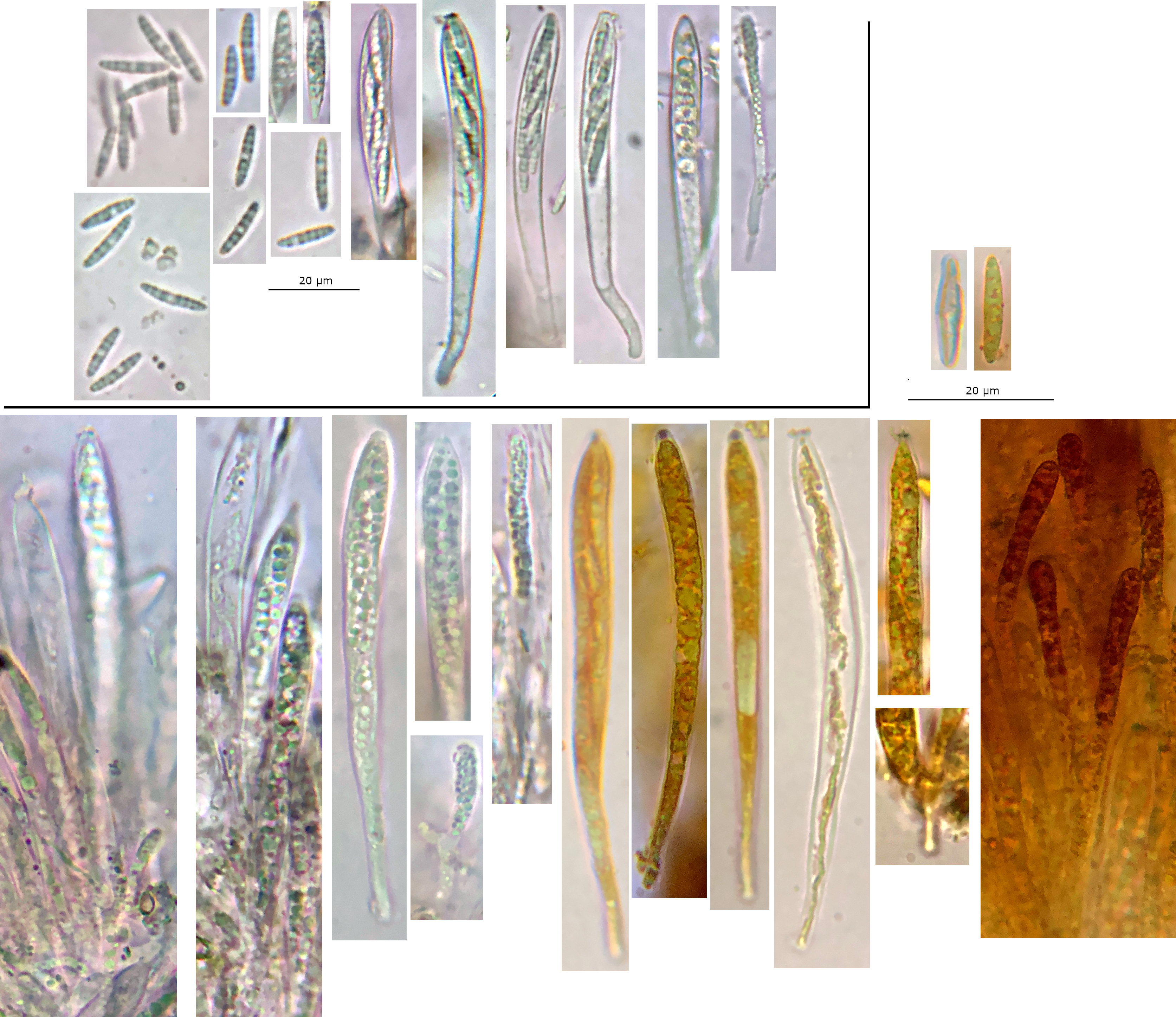
Asci: Cylindrical-clavate, apex hemispherical to conical-truncate, apical ring hymenoscyphus-type simple base – few seen were h-shape, 1-2-seriate, mostly breaking above the base so lengths may be underestimate.
- Vital: ~75-90 x 7-8 um, pars sporifera ~40%, mildly clavate, apex hemispherical when fully turgid, biseriate, often with two spores at the apex.
- Dead: ~65-80 x 5-6.5 um, pars sporifera up to 90%, more cylindrical, apex becoming acute-truncate to mammiform, apical thickening ~1-3 um, more uniseriate below.
- Discharged: Width ~5 um, cylindrical, apex lageniform with distinct neck and lip, apparently thickening fully everting and often not retracting – resulting in bumpy appearance of lip, usually still with amyloid material.
Spores: Ellipsoid-fusiform, almost all aseptate but one uniseptate and brownish (overmature), mostly homopolar tapering to rounded-acute poles, usually inequilateral, sometimes very slightly curved, many small VBs - OCI ~5 (vital), usually with a single whitish nucleus showing.
Vital spores in water: (12.9) 13.5-16.3 (25.4) × (2.5) 2.7-2.9 (4.2) µm, Q = (4.6) 4.8-5.8 (6.4), n = 15, mean = 15.7 × 2.9 µm, Q mean = 5.4. Several large spores ~24 x 4.2 um found outside asci, excluding these then mean = 14.5 x 2.8 um.
Paraphyses: Cylindrical, ~3-4 um wide, apparently 2 septate, many smallish VBs in upper part, dextrinoid contents.
VBs: In spores, paraphyses, cortical cells of ectal excipulum, reddening when traumatised, greenish at high magnification, appearing brown to brownish-rose in concentration and low magnification.
Exudate: Brownish-rose around margin and flanks, ectal excipulum gelatinised in some areas.
Medullary ex: Hyaline, textura intricata, separated from ectal excipulum by thick band of horizontal hyphae.
Ectal ex: Textura prismatica, areas of cortical cells with VBs forming lattice-like layer in surface view, with short protruding cylindrical hyphae/hairs at the stipe.
Marginal cells: Unremarkable, apical cells mildly clavate and elongated, and not protruding noticeably above hymenium.
Basal attachment: Not observed.
 Hymenium-0010.jpeg
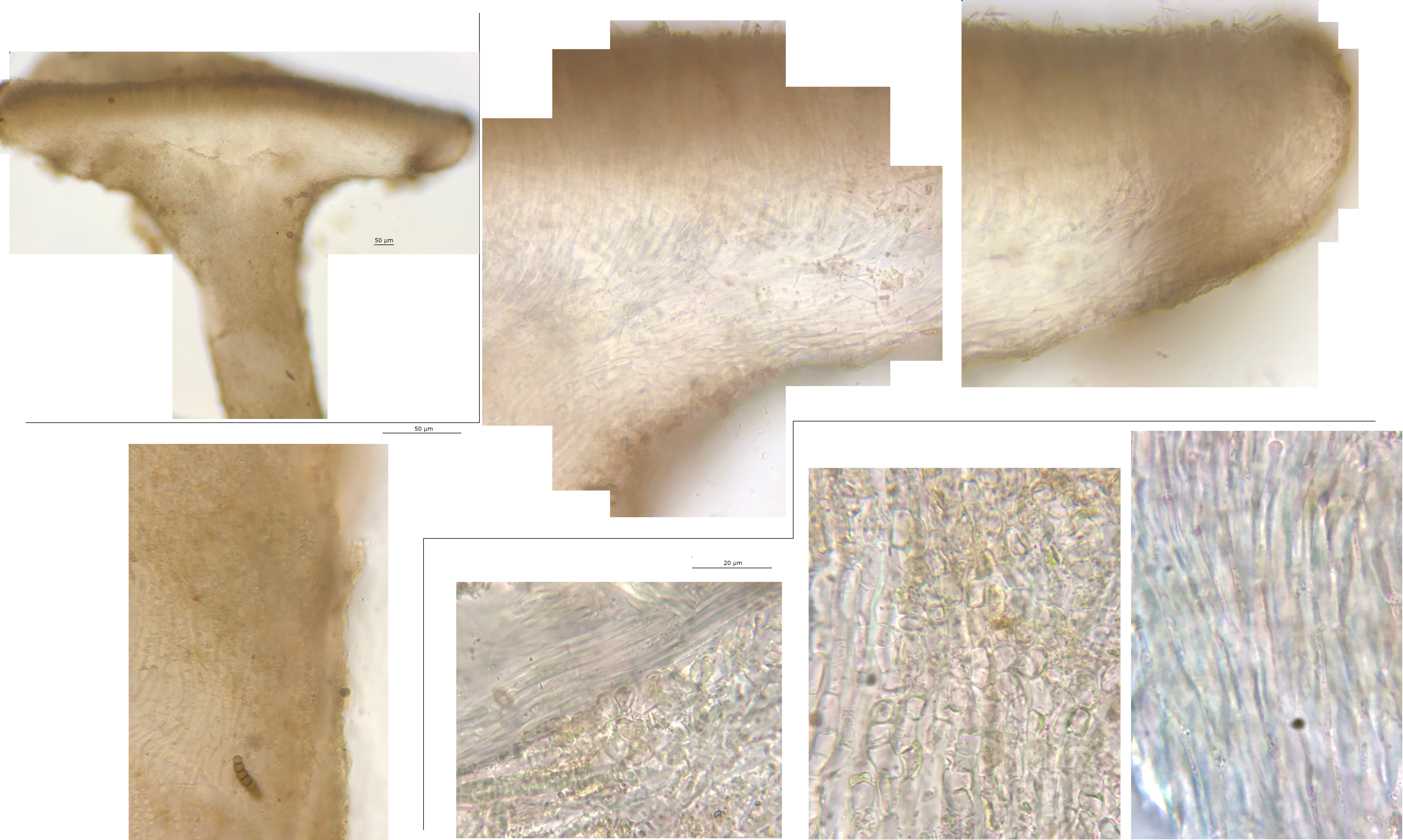 ExcipulumStipe-0001.jpeg
There are several similarities in micro with the few Hymenoscyphus spp I have seen, but I have not seen any Cyathicula spp before to understand the differences. Thanks for explaining about the rings, I will review the types.
Yes, C. paludosa (H-) does look to be right from the photos in your folder. The spores look identical, and I guess the bigger spores I saw are unrelated (especially as I didn't see any in mature asci). It also explains several other things such as the colouring, IKI reactions, the shape of the ascus apex, and the capitate hairs/protruding cortical hyphae on the ectal excipulum.
No, I meant the two vertically oriented spores that are > 20um long, to the right of the group of pictures of spores.